
#SPPC2023 - 6th September 2023, Lincoln
The 2023 Symposium on Palaeontological Preparation and Conservation will take place on 6th September 2023 at the University of Lincoln, UK
#SPPC2023 will take place at the Minerva Building, Lincoln UK, in conjunction with the Symposium on Vertebrate Palaeontology and Comparative Anatomy. Platform presentations will take place in the morning, with time during tea break and lunch for delegates to view posters.
For more information please contact
 Schedule
|
9:30am |
Registration (tea and coffee, posters)
|
|
|
10am |
Intro and Housekeeping
|
|
|
10:10am |
Frill seekers: using methyl cellulose to help remove plaster and burlap from Triceratops bone
|
by Kieran Miles
|
|
10:30am |
An Introduction to Solvent Gels in the Conservation of Geological Materials
|
by Lu Allington-Jones
|
|
10:50am |
Please Handle with Care: The Conservation of Highly Fragmented Fulgurite and its Display.
|
by Efstratia Verveniotou
|
|
11:10am |
Break (tea, coffee, posters) |
|
|
11:40am |
How to become a fossil preparator in Europe? |
by Cinzia Ragni and Marina Rull |
|
12:00noon |
Time for an update: Diprotodon, giant wombat-like, over 100 years of history, on display at the Natural History Museum.
|
by Efstratia Verveniotou, Spyridoula Pappa, Ellie Clark and Lauren Burleson
|
|
12:20pm |
Unravelling the Mysteries of the Megalosaurus Moulds |
by Emma Nicholls*, Juliet Hay, and Eliza Howlett |
|
12:40pm |
Finish |
|

#SPPC2023 - Abstracts

Poster Abstracts
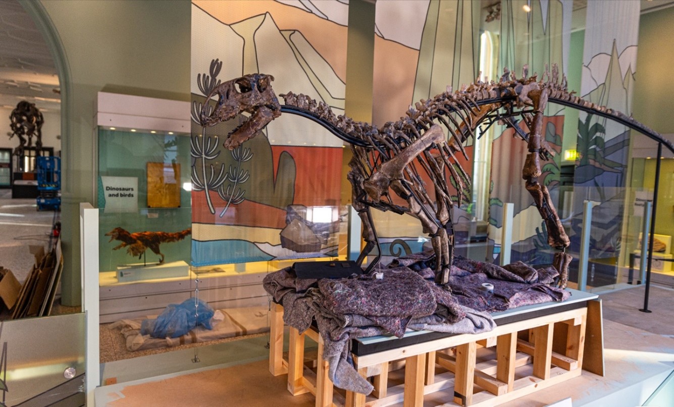
The Conservation and Installation of a Tenontosaurus Skeleton at the Manchester Museum
Jenny Discombe1 and Emily Gershman2
1Manchester Museum UK; 2Durham University
In 2004, a Tenontosaurus skeleton, known as April, was taken off display after research indicated that it had been mounted inaccurately. 19 years of conservation and academic research has culminated in April finally being re-exhibited this past February. The primary aim for the conservation and redisplay of April was to present the skeleton honestly and accurately without the use of excess filler or replicas common in most dinosaur displays, and to ensure it remained accessible for future research. To achieve this goal, aged adhesive and filler were mechanically removed and any glossy varnish was cleaned from fossils using a laser. The fossils were repaired using an appropriate adhesive and small amounts of an epoxy putty where necessary. The long bones were strategically left broken as their cross sections have potential for future research. To mount the specimen, a custom frame was commissioned which could be manipulated to support each bone in an anatomically accurate position. Fishing line, in conjunction with various barrier materials, was then used to attach the fossilised bones to the frame. The result of this approach is less visually obtrusive when compared with traditional mounts, it allows for easy removal of bones for research, it enables more flexibility in the positioning of the bones to create a more anatomically accurate display, and it presents a unique opportunity to see the variable preservation levels in fossilized dinosaurs.
Radioactive fossils: What? Where? Why? - and how to reduce the risk to health
Nigel Larkin1 & Jana Horak2.
1 School of Biological Sciences, University of Reading, Whiteknights, Reading, RG6 6EX, UK.
2National Museum Wales, Cardiff CF10 3NP, UK.
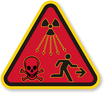
Most curators and collection managers responsible for a geological collection will be aware of the risk posed by rocks and minerals which emit ionising radiation and will have put measures in place to reduce the risk to collection users. However, fewer people are aware that fossils can be radioactive too, from fish to dinosaurs – even coprolites. A fossil may be radioactive if it has been exposed to uranium-bearing fluids during diagenesis and fossilisation processes. Any groundwater moving from the radioactive deposit to or through another nearby porous deposit can contaminate the rocks and fossils with radioactive elements. Over time the skeletons or other remains being fossilised incorporate these elements into their structure. The processes of fossilisation can even concentrate the levels of radioactive elements so that some fossils may be more radioactive than their host rock. The more massive the fossil, e.g. a large dinosaur bone, the greater the potential for a higher level of radioactivity, and a greater risk to humans. Whilst most radioactive fossils may not present a danger to health through direct absorption of radiation unless there is prolonged exposure at close quarters (e.g. hundreds of hours), the main risk is posed by the ingestion or inhalation of small radioactive particles from the specimen. Here, we describe what measures can be taken to reduce these risks and also provide a list of locations around the world known to yield radioactive fossils which we hope can be augmented by others.
The technique of preparation and conservation of samples from microvertebrates collecting from the upper Miocene of Romania
Dumitru-Daniel BADEA1*, Bogdan-Gabriel RĂȚOI1, Mihai BRÂNZILĂ1
1Universitatea “Alexandru Ioan Cuza” din Iaşi , Bulevardul Carol I, Nr. 20A, Corp B, Iaşi-700506
*e-mail:
Fossil elements belonging to microvertebrates are often identified after screen-washing the studied sediments, using stable sieves. Usually the smallest sieve has meshes of 0.063 mm. Binocular magnifiers are used to identify the fossil material, but the identification is not precise and it is necessary that the fossil elements be prepared in order to photograph them with high-quality microscopes, such as digital microscopes or electronic microscopes. Electron microscopy highlights the smallest morphological details of fossil elements, which helps to identify them at the species level. The most important fossil elements identified in the upper Miocene of Romania are teeth of microvertebrates, fish, reptiles, rodents, insectivores, lagomorphs or even fossil bat teeth. Besides these, gastropods, eggshell fragments or postcranial bones were also identified here.
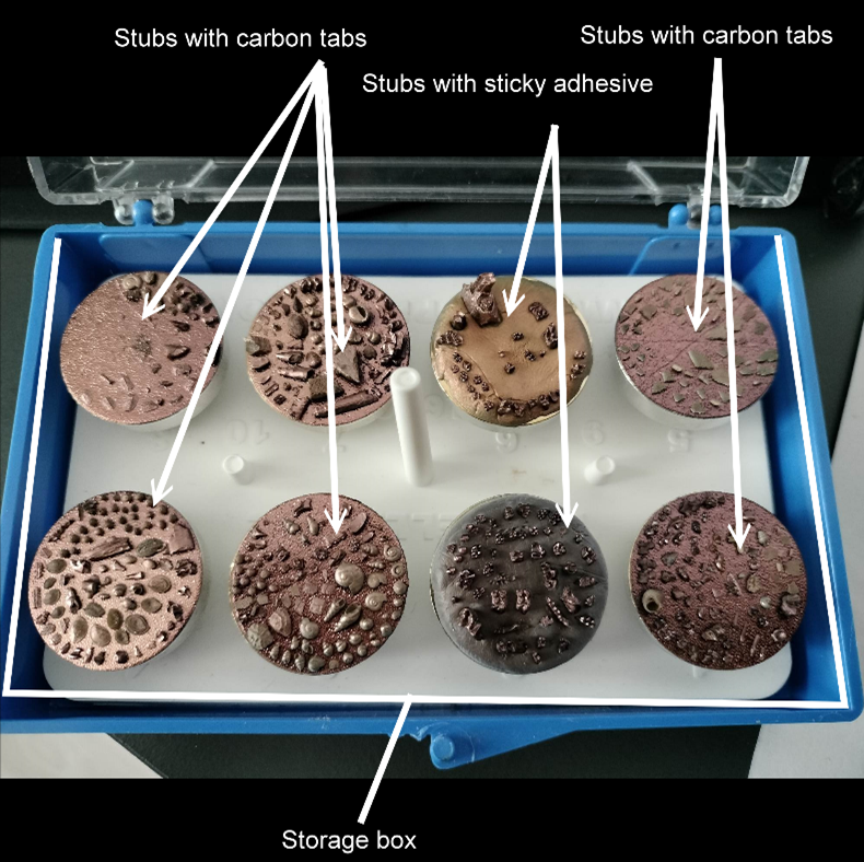
All these fossil elements were processed to be photographed with the SEM microscope, model Hitachi S-3400N II. Hitachi stubs of Ø25mm were used over which it sticks the high purity conductive double sided adhesive Ø25mm carbon tabs making it easy to attach elements with at least one flat surface (gasteropods, teeth without roots, osteoderms etc.). Fossil teeth with roots were fixed in professional sticky adhesive and then positioned on stubs thus making it possible to analyze their occlusal part. Prior to the the photography process, the surface of the fossil elements was coated with an electrically conductive thin film (14 nm gold) to inhibit charging and enhance secondary electron emission. This thin film was deposited using a versatile Q150T S sputter coater (Quorum Technologies) fitted with a film thickness monitor that allows control of the coating applied to the samples. Stubs of this type can host at least 50 elements of microvertebrates, facilitating the conservation process of small fossil elements. All the stubs are grouped by eight in a storage box (see Fig.1). Thus, the small fossil elements can be kept for possible new investigations and analyses, easily exhibited in the museum exhibitions where they will be recorded.
Investigating alternative materials for fossil mounts
Arianna Bernucci and Erica Read, The Natural History Museum, London UK
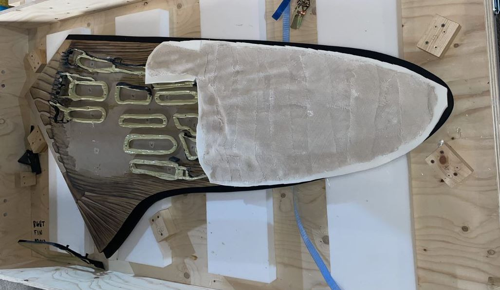
The conservation department at the Natural History Museum (London) has traditionally used Epopast 400 (two part epoxy and fibreglass resin) to create strong supportive mounts for fossilised specimens. Epopast can be moulded to any shape and can be used with heavy or delicate specimens, while providing an even weight distribution thereby making the specimens more accessible and safer to handle. Although these mounts are strong and reliable, their fabrication is resource heavy and are not without significant health and safety risks. Two alternative materials have been found that may be suitable replacements - EMP60, a glass-filled epoxy laminating/reinforcement paste, and AquaResin, a 2-part polymer emulsion with a compatible gypsum derivative. Both materials were assessed for curing and processing times, shrinkage, tensile strength, ease of handling, cost and health and safety risks with promising results.
Wild Ox on the Move: conserving and increasing accessibility of fossil aurochs found in the Uphall Brickfield at Ilford, Essex in 1864 ahead of a collection moves project
Lucia Petrera*, Fabiana Portoni*, Erica Read*, Lauren Burleson*, Spyridoula Pappa*,**
*Natural History Museum, London UK
**Royal Holloway University of Londonas been the focus of a conservation and restorage project as part of the NHM Unlocked Moves Programme.
Aurochs were a giant, wild ancestor of modern cattle. They first appeared around half a million years ago and spread across Europe, Asia, and North Africa. The bulls were nearly two metres tall. The fossils in this project are oversized, heavy, fragile and required a highly specialised approach to ensure their safe transportation and long-term storage and preservation.
The project began with a comprehensive condition assessment, which identified areas of damage and deterioration. A team of conservators subsequently completed a range of treatments (including cleaning, consolidation, and repair of damaged areas) to ensure specimens’ stability. Following storage assessment, bespoke foam-lined epoxy resin mounts (Epopast 400 ‘jackets’) with aluminium board were built enabling protection during transfer and to protect and secure the future of this collection.
Bespoke Epopast 400 jackets have proven to be an effective solution for supporting the uneven weight distribution of the specimens. They increase accessibility to important collections for upcoming future research projects and improve their long-term preservation as well as stability during transportation.

X-Ray Diffraction as a tool for assessing the effect of conservation treatments on sub-fossil bone
Lu Allington-Jones, The Natural History Museum, London UK
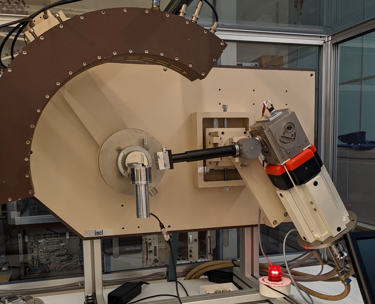
Conservation treatments which aim to clean, consolidate or adhere sub-fossil bone must be chosen with consideration for their potential to affect preservation and research data. XRD analysis was investigated in this study to evaluate its potential for detecting crystallographic differences in sub-fossil bone samples following treatment with common conservation chemicals. Nineteen remedial treatments are tested to identify high and low risk treatments to guide conservation decision-making and enable more-informed treatment plans. The study concludes that XRD analysis can successfully be used to evaluate conservation treatments but should not be considered in isolation.
Platform Presentation Abstracts
Time for an update: Diprotodon, giant wombat-like, over 100 years of history, on display at the Natural History Museum.
Efstratia Verveniotou1, Spyridoula Pappa1, 2, Ellie Clark1 and Lauren Burleson1
1Natural History Museum, London
2 Royal Holloway University of London
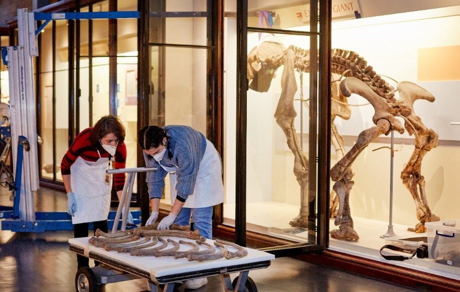
The Natural History Museum houses a mounted Diprotodon skeleton, which first arrived in London in 1907, its rich history stretching back over 100 years. After many years on display the specimen was completely disassembled, moved, conserved, and reassembled as part of an extensive 2022 Gallery Refurbishment Project, aiming to secure the future of this historically important specimen for another 100 years.
Diprotodon was an enigmatic species to European science, until described by Richard Owen in 1838, and is the largest known marsupial to have existed, living during the Pleistocene (2.6mya to 11,700 years ago) in Australia. During the refurbishment project scientists discovered historically important photos and publications in link with the specimen from the Museum Archives. Its full reconstruction was completed by the prominent Australian zoologist, Dr E.C. Stirling and Mr Zietz in the South Australian Museum in 1905. They included limb and caudal vertebrae fossils from Lake Callabonna, a site of exceptional fossil preservation in South Australia, and based the rest on specimens from Adelaide and Queensland fossil sites.
The 103 skeletal elements had been mounted in an intricate Victorian mount through an interlocking technique and use of wire. The mount had started to corrode in places, cutting through the bones risking damage to the specimen. The original mount was kept but upgraded considering current conservation practices which facilitated the safe re-display of delicate bones. The installation of the specimens inside its display cases presented a series of technical difficulties that took several museum specialists and equipment to address.
Frill seekers: using methyl cellulose to help remove plaster and burlap from Triceratops bone
Kieran Miles, Natural History Museum, London UK
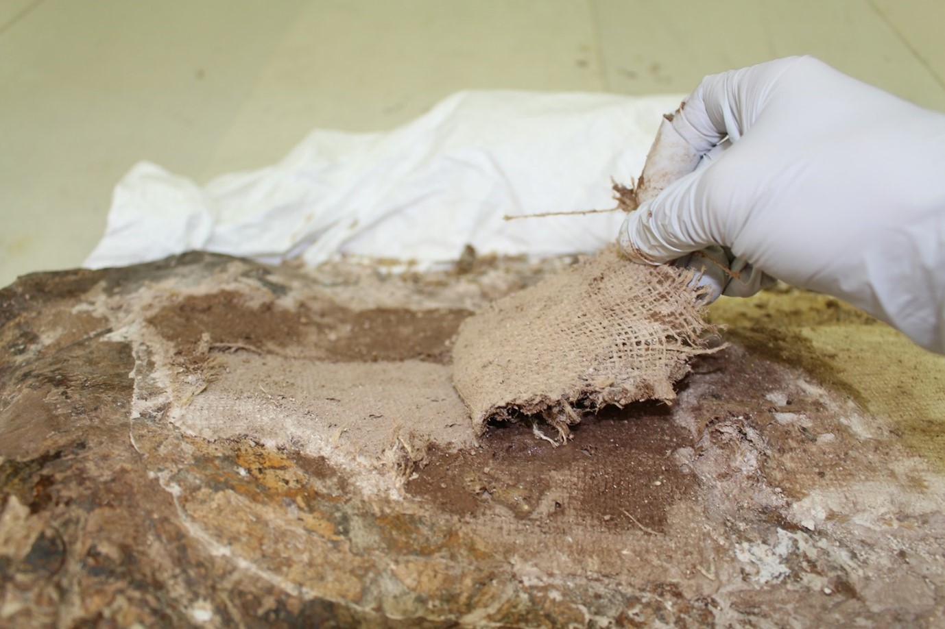
The dinosaur collection at the NHM includes a Triceratops skull (NHMUK PVR3886), purchased from Charles Sternberg in 1909. While most of the skull is on public display in the Dinosaurs gallery, two large sections of the frill (the squamosal bones) had been replicated for the display and the originals left in storage. With much of the fossil bone obscured by plaster of Paris, the original frill sections were deemed of limited use to science in their current form. A work request was received to strip away as much artificial material as possible to determine the extent of the original fossil material. An air scribe fitted with a chisel stylus proved initially effective, but soon revealed an additional obstacle – layers of hessian underneath.
The use of a methyl cellulose poultice was trialled to swell the hessian, aiding softening of the plaster, and allowing layers to be torn off in strips. The methyl cellulose was applied by paintbrush, then covered with cling film and left for 1 hour. It was then uncovered, and a metal modelling tool was used to lift the softened layers of hessian. The process was repeated until most of the plaster and hessian had been removed. The fine layer of plaster remaining on the bone was removed, where possible, with a Split-V ultrasonic tool. This sequence of treatments can be utilised at minimal risk to specimens in similar situations when a hessian and plaster jacket has been applied but a separator layer has been omitted.
Please Handle with Care: The Conservation of Highly Fragmented Fulgurite and its Display.
Efstratia Verveniotou, Natural History Museum, London UK
This article presents the conservation treatment of a highly fragmented thin tube of natural silica glass formed when a lightning strike the sandy dunes of Maldonado, Uruguay. The fulgurite which would have been almost one meter long was broken in eight main pieces with numerous sandy friable smaller parts. The specimen had evidence of being adhered together in the past and presented as a complete piece in a bespoke wooden box. Its old adhesive was yellowing, brittle and failing. Internally the fulgurite was thin smooth glass and externally sandy, friable and porous.
The aim of the conservation treatment was to fully reconstruct the specimen and redisplay it vertically in its historic box. The advantages, disadvantages, and ethical considerations of using replacement material are discussed along the testing of different conservation materials considered. The chosen methodology allowed the full reconstruction of the fulgurite as all fragments were placed back on the specimen. A bespoke mount allows the specimen to be safely displays in any orientation necessary while respecting the historic character of its wooden display box.

An Introduction to Solvent Gels in the Conservation of Geological Materials
Lu Allington-Jones, The Natural History Museum, London UK
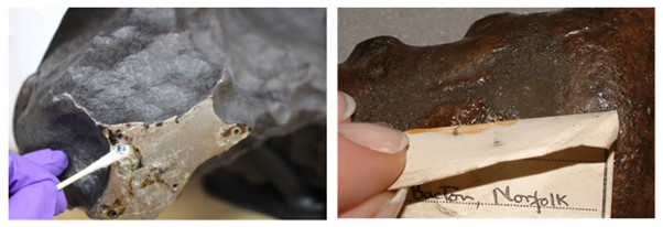
Solvent gels comprise a solvent or solvent mixture thickened by a gelling agent. Their high viscosity enables more control and reduces contamination of a substrate compared with fluid solvents. They also have health and safety advantages. Solvent gel recipes can be tailored to hold different solvents, to different consistencies and also to carry other chemicals such as chelators. Examples of use on geological collections include resin and iron oxide removal, label detachment and the controlled delivery of ethanolamine thioglycolate for the treatment of pyrite oxidation.
How to become a fossil preparator in Europe?
Cinzia Ragni1 and Marina Rull2
1UNITO, University of Turin, Via Valperga Caluso, 35, 10125 Torino TO, Italy
2Conflent Spéléo Club de Prades, 4, Travers de Fabriques, Prades de Conflent (Occitane, France).
The preparation of fossils is the necessary first step to permit the future study and exhibition of fossil specimens. However, do specific university studies in it exist? Did all the current fossil preparators from European countries follow the same formative path?
 |
Unfortunately, fossil preparators who work at European institutions followed different academic itineraries or studies, in most cases according to the different traditions or specific legislations of each country. A consensus on how to become a fossil preparator does not seem to exist, nor official university studies focused on it in most countries in Europe. Fossil preparation needs knowledge of biological, geological and chemical sciences, as well as expertise in restoration and conservation procedures. This multi-disciplinary profile is quite difficult to find in one single degree programme. Consequently, in reality, fossil preparators become experts in their field thanks mostly to their professional experience and through multiple unofficial formative courses. It is also worth mentioning the existence of another type of freelance preparator, not permanently linked to any research/exhibition institution, who customarily work and prepare fossils for sellers. These commercial preparators usually have a more artistic approach according to the seller’s demands as compared with preparators linked to scientific institutions, whose approach is based always on the extraction of scientific information. In Italy, where not all museums have the possibility to create their own laboratory, they ask for the support of preparators to do preventive conservation, preparation, modelling, exhibition etc. In this presentation, and on the basis of the experience of different institutions in different countries (like Italy, Spain, Germany, Switzerland and others), we will discuss the path to becoming a fossil preparator, the present possible ways and future perspectives. |
Unravelling the Mysteries of the Megalosaurus Moulds
Emma Nicholls*, Juliet Hay, and Eliza Howlett
Oxford University Museum of Natural History
Megalosaurus (Buckland, 1824) is famous for being the first dinosaurian taxon to have been scientifically described. It is also one of three genera upon which Richard Owen based the name Dinosauria, in 1842. Subsequently, with regard to the history of science at least, it could be argued that Megalosaurus is one of the most important dinosaurs in the world.
The first Megalosaurus fossils known were found in Stonesfield Quarry, Oxfordshire, in the late 18th and early 19th centuries. Some of the material was subsequently acquired by W. Buckland and described in a paper to the Geological Society of London as Megalosaurus, in 1824. The specimens included a femur, ribs, vertebrae and a right dentary, all of which were illustrated in Buckland’s paper by his future wife Mary (née Morland). With no holotype, the right dentary (OUMNH PAL-J.13505) was declared the lectotype (Molnar, 1990), thus solidifying its place in the history books as the type specimen of the first scientifically described dinosaurian taxon.
It is thought that over the last two hundred years, replicas of this extremely important specimen have been made using techniques from moulding to 3D printing. Research into what exists, how they were made, and where they are housed, forms the basis of this paper, from the historical cast made by (or on behalf of) W. Buckland, to the latest 3D render, printed with dark teeth and transparent bone to show internal structures.
Presentations from previous years can be viewed here
For more information please contact


